

In the medical field, radiology holds a dominant position. It is utilized for advanced testing and treatment, disease detection, wellness checkups as well as screening. Here these methods are used, as it aids the medical professionals to see inside the body and then diagnosing and treating it. As technology is advancing rapidly, the methods inputted in these fields are also advancing with time.
What is Radiology?
“Radiology is essential to the diagnosis of many diseases, particularly cancer. Early diagnosis saves a life. Without diagnosis there can be no treatment, there can be no cure” – Ontario Association of Radiologists
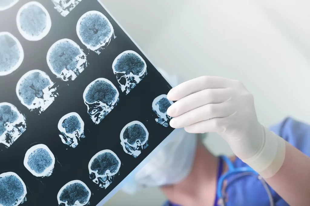
Radiology is considered to be the most technologically advanced field of medicine. Radiology is the medical discipline that makes use of medical imaging to identify illness and direct treatment in the bodies of humans and other animals. Radiology is also known as diagnostic imaging, where a series of tests are conducted which take the images of various parts of the body to examine.
A diagnostic imaging centre has the equipment and trained specialists to perform the tests. The imaging equipment used in diagnostic imaging centres are many and depending on the requirement of the patient either they are used separately or combined. There are many different imaging tests and among them, the popular ones are X-Ray and MRI. Ultrasonography and Magnetic Resonance Imaging(MRI) don’t use electromagnetic radiation while Computed Tomography(CT), Fluoroscopy and nuclear medicine including Positron Emission Tomography(PET) employ it.
There are three different types of radiology:
- Diagnostic Radiology- This category of radiology allows medical professionals to view structures inside your body with the help of images. These images help the doctor to understand the cause of your symptoms, keep track of how well your body is responding to the treatment and check for diseases like cancer and heart diseases.
The most common diagnostic radiology tools are- CT Scan, MRI, Magnetic Resonance Angiography(MRA), Mammography, Ultrasound and X-Rays.
- Interventional Radiology- This is a specialized field in radiology as here doctors undertake minimally invasive surgical operations using tiny incisions in the body in addition to interpreting your medical images. According to professionals in this field, it isn’t always the best procedure for your condition, but it is more effective than traditional treatments. This category offers benefits such as lower costs, more comfort and faster recovery.
- Therapeutic Radiology- Also referred to as Radiation Oncology or Radiation Therapy, this is a practice of using radiation to treat cancer and other diseases. Here radiation is used to destroy cancer cells as well as stop them from growing or spreading further. Cancer symptoms can be reduced or treated by Therapeutic radiography. Treatments are done either alone or in conjunction with chemotherapy and other treatment approaches.
Since its discovery in 1985, radiology has gradually grown to become an important medical speciality. The versatility of imaging technology has cemented radiology’s place in medical care. Currently, radiology serves as an important diagnostic tool for a wide range of diseases. It aids in treatment monitoring and can even forecast treatment outcomes. With a variety of technologies and procedures available for the diagnosis, staging and treatment of diseases, radiology plays a key role in disease management. The detailed information provided by diagnostic imaging gives an overview of the structural or disease-related changes. Early detection saves a life. So thus without a diagnosis, there can be neither therapy nor cure.
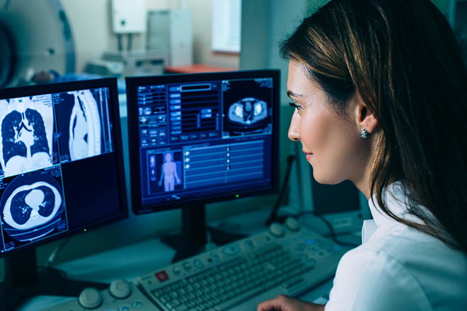
What do radiologists do? The American College of Radiology defines radiologists as “ medical doctors(MDs) or doctors of osteopathic medicine(DOs) who specialize in diagnosing and treating injuries and diseases using medical imaging (radiology) procedures (exams/tests) such as X-rays, computed tomography (CT), magnetic resonance imaging (MRI), nuclear medicine, positron emission tomography (PET) and ultrasound.”
The main functions of radiologists include:
- Deciding which imaging test is most appropriate for your needs
- Examining and analyzing the medical images from your examination.
- Provide the physician with a detailed diagnostic report of their results.
- Instruct the radiology technicians about operating the imaging devices during the test.
DIFFERENT DIAGNOSTIC IMAGING TESTS INCLUDE:
X-RAYS–
X-rays are a type of radiation known as electromagnetic waves and this is the most commonly used diagnostic imaging test. X-ray imaging produces images of the inside of your body. The images depict different parts of your body in various shades of black and white. This is because different tissues absorb different amounts of radiation. It is performed for many reasons which include understanding the cause of pain, monitoring the progress of a disease, and for evaluating how well a treatment is working. X-rays involve directing a small amount of radiation to the desired location of the body where images are required for the process.
A Computed Tomography scan which is also referred to as CAT(Computed Axial Tomography) scan allows doctors to view cross-sections of the body. A CT scan is a diagnostic imaging procedure that creates images of the inside of the body using X-rays and computer technology. It displays high-resolution images of any part of the body, including the bones, muscles, fat, organs, and blood vessels. CT scan provides more detailed information than X-rays. CT scans may be used to assist in the diagnosis of tumours, investigate internal bleeding, or look for other internal injuries or damage. Tissue or fluid biopsy can also be performed using CT.
How long do CT Scan results take? The results of the scan usually take 24 hours. A radiologist, a physician who specializes in reading and interpreting CT scans and other radiologic images, will review your scan and prepare a report that explains it. In an emergency setting, such as a hospital or emergency room, healthcare providers frequently receive results within an hour. After a radiologist and your healthcare provider have reviewed the results, you will either have another appointment or receive a call. Your healthcare provider will go over the results with you.
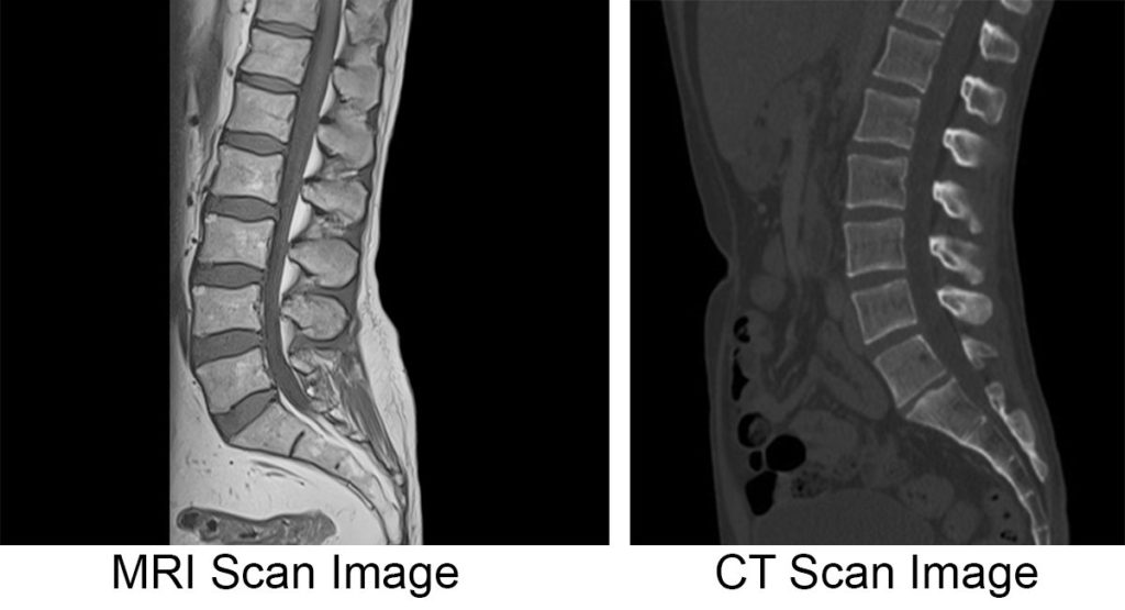
X-RAYS VS CT SCAN:
CT scans provide more information than standard X-rays. A beam of energy is directed at the body part being studied in standard X-rays.
X-Rays – Creates 2D images
Typically used to view and diagnose fractures, dislocations,
infections and tumours
It is the most common and widely available test
Use radiations to produce images
CT Scan – Creates 3D images
It produces detailed images of organs, bones, soft tissue, and
blood vessels
More powerful than X-Rays
Take a 360-degree image
MRI–
Magnetic resonance imaging (MRI) is a medical imaging technique used in radiology to create images of the body’s anatomy and physiological processes. MRI scanners generate images of the organs in the body by using strong magnetic fields, magnetic field gradients, and radio waves. The majority of MRI machines are large, tube-shaped magnets. When you lie inside an MRI machine, the magnetic field temporarily realigns the water molecules in your body. Radio waves cause these aligned atoms to emit faint signals, which are used to generate cross-sectional MRI images. The major advantage is that no ionizing radiation is used.
Difference between MRI AND X-Rays? X-rays are more quickly available than MRI images and can be used to diagnose injuries and masses inside the body. MRIs may provide clearer, more detailed images of tissues and organs such as the brain. X-Ray is relatively cheaper than MRI.

SONOGRAPHY –
Sonography is a technique that produces images of internal body structure by using high-frequency sound waves. Sonography is also known as ultrasound and it is a non-invasive, painless procedure. Ultrasound waves are used for this purpose. Images of organs, tissues, and blood flow are created. A sonogram is an image produced by ultrasound. Ultrasonography is another name for sonography. A small device called a transducer is placed against the patient’s skin near the body area to be imaged during the sonographic process. Because it can transmit and receive sound, the transducer functions similarly to a loudspeaker and microphone.
TYPES OF ULTRASOUND;
- Endoscopic ultrasound.
- Doppler ultrasound.
- Color Doppler.
- Duplex ultrasound.
- Triplex ultrasound (colour-flow imaging)
- Transvaginal ultrasound.
MAMMOGRAPHY–
Mammography is a type of medical imaging that uses a low-dose x-ray system to examine the inside of the breasts. A mammography exam, also known as a mammogram, helps women detect and diagnose breast diseases early. Mammograms, like all X-rays, use ionizing radiation to create images. These images are then examined for abnormal findings. Lower-energy X-rays, typically Mo (K-shell X-ray energies of 17.5 and 19.6 keV) and Rh (20.2 and 22.7 keV) are typically used more than those used for bone radiography. Digital mammography, computer-aided detection, and breast tomosynthesis are three recent advances in mammography.
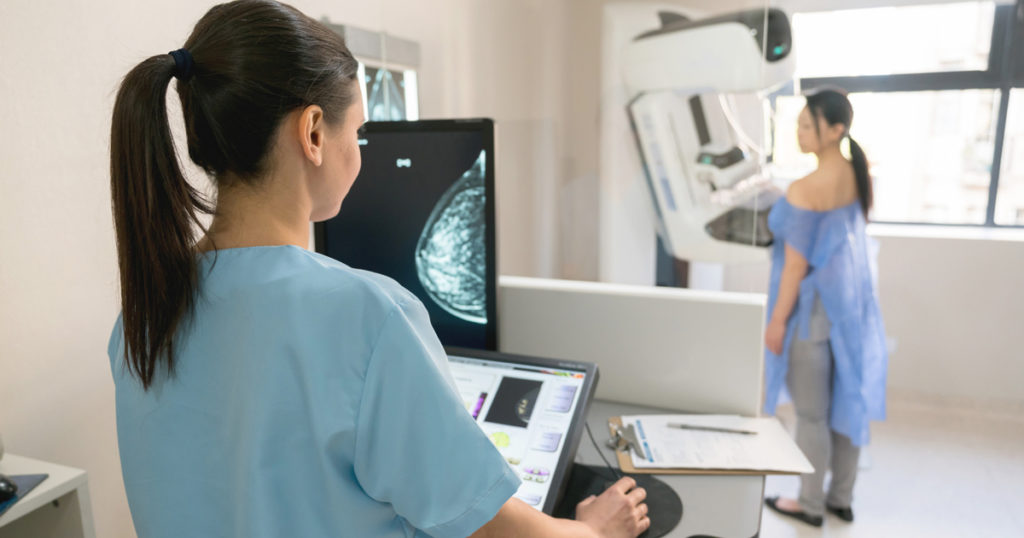
What is a diagnostic mammogram?
A diagnostic mammogram is used to look into suspicious breast changes like a new breast lump, breast pain, an unusual skin appearance, nipple thickening, or nipple discharge. It’s also used to evaluate abnormalities on a screening mammogram. Additional mammogram images are included with a diagnostic mammogram.
The patient can benefit from radiology in a variety of ways. Radiology benefits patients by using advanced tools, techniques, and treatment options to detect and treat disease. Diagnostic imaging facilitates the acquisition of detailed information about disease-related changes or structural variations. It enables the patient to detect problems early, increasing their chances of healing themselves. Saving lives would be difficult without radiology. Medical imaging is critical for treating and diagnosing severe conditions. Without the process, the number of people who die will continue to rise.
So in this article, detailed information about radiology and different types of it is provided. The article also elucidates the importance of radiology and different diagnostic imaging tests. In the next article, more diverse information regarding radiology will be discussed and clarified. Until then stay in touch with us and I hope you are healthy and safe.