

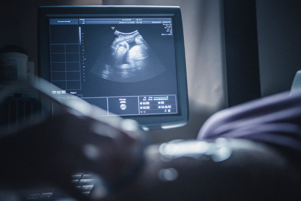
If you have never had an ultrasound check in your life. however, you would like to understand how it works. Then let me help you to know everything concerning Ultrasound scans. If you watch loads of films then, you need must have heard the term “ultrasound”, doubtless in every scene wherever a pregnant girl in her doctor’s workplace gets a sneak peek of the baby growing within her female internal reproductive organ — even perhaps sorting out whether or not they ought to paint the nursery pink or blue. However whereas fetal imaging is one of the foremost common uses of ultrasounds, this diagnostic tool truly has several applications.
What is Ultrasound Imaging and How it Work?
Ultrasound imaging uses high-frequency sound waves to make a picture of the within of your body. It’s excellent at observing the soft tissues of the body and is commonly the primary step in deciding the cause for your symptoms.
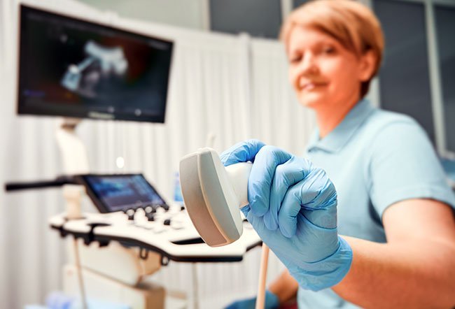
Also called ultrasonography, ultrasound imaging uses a little electrical device (probe) to transmit sound waves into the body and record the waves that echo back. Sound waves travel into the realm being examined till they hit a boundary between tissues, like between fluid and soft tissue, or soft tissue and bone. At these boundaries, a number of the sound waves are mirrored back to the probe, whereas others travel any till they reach another boundary and are mirrored back. Since the speed, direction, and distance sound waves travel dissent betting on the boundary they run into, a laptop will interpret this data as a two-dimensional image on a screen.
The shape and intensity of the echoes depend upon however the realm absorbs the sound waves. For instance, most waves go through a fluid-filled cyst and challenge only a few or faint echoes that look black on the video display. On the opposite hand, waves can bounce off a solid neoplasm, making a pattern of echoes that the pc can interpret as a lighter-colored image. Air and bone additionally replicate sound waves.
Risk Factors-
It has been around for over sixty years and is taken into account safely since there are not any better-known risks and it doesn’t use radiation. It’s one among the foremost usually ordered imaging exams since it’s versatile, portable, comparatively cheap, non-invasive, and may give time period data concerning the realm of concern.
In short, ultrasound could be a safe procedure that uses low-power sound waves. There are not any better-known risks until this date.
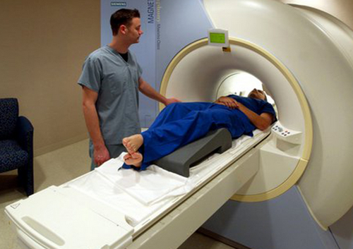
We all can trust the very fact that it could be a valuable tool, however, it’s limitations. Sound doesn’t travel well through air or bone, this ultrasound is not effective at imaging body elements that have gas in them or are hidden by bone, like the lungs or head. to look at these areas, your doctor could order different imaging tests, like CT or imaging scans or X-rays.
Uses of Ultrasound Tests-
- Ultrasound is employed for several reasons, together with to:
- View the womb and ovaries throughout gestation and monitor the developing baby’s health
- Diagnose vesica malady
- Evaluate blood flow
- Guide a needle for diagnostic assay or neoplasm treatment
- Examine a breast lump
- Check your ductless gland
- Detect reproductive organ and prostate issues
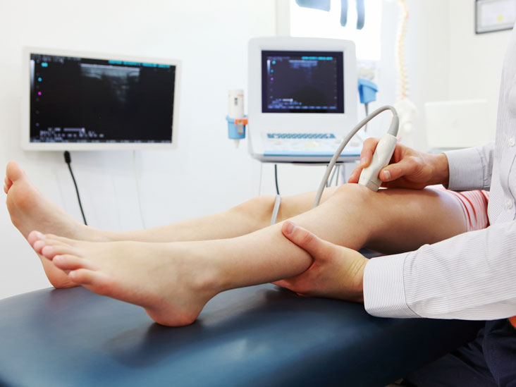
- Assess joint inflammation (synovitis)
- Evaluate metabolic bone malady
Types of Ultrasound
Most ultrasounds are done employing an electrical device on the surface of the skin. Sometimes, however, doctors and technicians will get a higher diagnostic image by inserting a special electrical device into one among the body’s natural openings:
In a transvaginal ultrasound, an electrical device wand is placed in a very woman’s epithelial duct to urge higher pictures of their womb and ovaries.
A transrectal ultrasound is typically employed in the identification of prostate conditions.
A transesophageal sonogram uses the electrical device probe within the esophagus in order that the sonographer will get clearer pictures of the center.
Additionally, ultrasound technology has advanced to permit various sorts of imaging:
- Doppler could be a special variety of ultrasound that makes pictures of blood flow through vessels.
- Bone ultrasonography helps doctors diagnose pathology.
- Echocardiograms are wont to read the center.
- 3D imaging adds another dimension to the ultrasound image, making three-dimensional interpretations instead of the flat two-dimensional pictures that are created with ancient ultrasound.
- 4D ultrasounds show 3D pictures in motion.