

Definition, Uses, procedure & Side effects: What is an echocardiogram used to diagnose? An echocardiogram is an ultrasound of the heart that your doctor uses to diagnose heart conditions. It uses sound waves to create pictures of your heart. This common test allows your doctor to have a better look at your heart condition like beating and pumping blood. Your doctor will use the photographs from the associate echocardiogram to spot your heart condition.
Why it’s done
Your doctor could recommend an associate echocardiogram to:
- Check for issues with the valves or chambers of your heart
- Check if heart issues are the reason behind symptoms like shortness of breath or pain in your chest.
- Detect inherent heart defects before birth (fetal echocardiogram)
- The type of echocardiogram you have got depends on the knowledge your doctor desires about your symptoms.
“Or to explain it in the shortest way we can call An echocardiogram, or “echo”, as a scan that investigates any issues with your heart by producing medical images of your heart with the help of ultrasounds.”
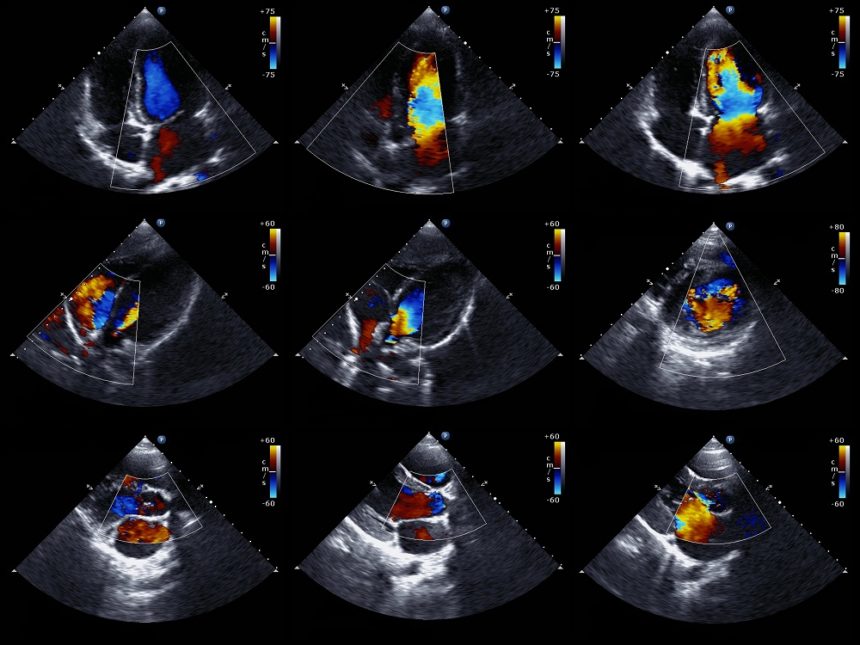
It’s a variety of ultrasound scans, which suggests a little probe is employed to send high-frequency sound waves that create echoes once they bounce off completely different components of the body.
This echoes and is picked up by the probe and changed into a moving image on a monitor whereas the scan is administered.
An echocardiogram could also be requested by a heart specialist or any doctor to confirm whether you have any heart condition or think you would possibly have a haul along with your heart.
The test will be performed at a hospital or clinic by a heart specialist or a trained specialist referred to as a cardiac physiologist.
Although it’s the same name, AN echocardiogram is completely different from AN graphical record, conjointly referred to as EKG, which may be a take a look at wanting to check the rhythm and electrical activity of the center.
What does an Echocardiogram show?
An echocardiogram will facilitate diagnosis and monitor sure heart conditions by checking the structure of the center and close blood vessels, analyzing how blood flows through them, and assessing the pumping chambers of the center.
An echocardiogram will facilitate discovering the subsequent conditions.
Damage from an attack because of a blockage within the coronary arteries, compromises the blood flow to the center muscle.
- Heart failure – wherever the center fails to pump enough blood throughout the body PRN.
- Congenital heart condition – birth defects that have an effect on the conventional functions of the center.
- Problems with the center valves- like a rheumatic heart condition.
- Cardiomyopathy – wherever the center walls become thickened or enlarged, compromising the pumping action of the center.
- And Endocarditis– AN infection of the center valves.
An echocardiogram is additionally facilitated to decide the precise treatment for an exact condition.
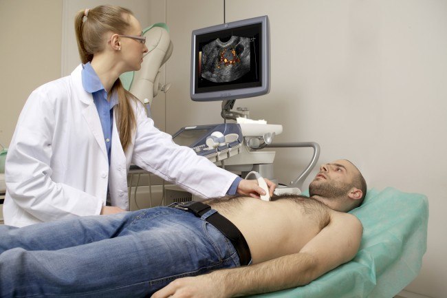
There are many alternative ways AN echocardiogram may be administered, however, the general public can have a transthoracic echocardiogram or TTE.
You won’t typically have to be compelled to do something to arrange for the take a look unless you are having a transesophageal echocardiogram.
For a TTE, you will be asked to get rid of any covering your higher 0.5 before lying down on a bed. you will be in a hospital suite to hide yourself throughout the take a look.
When you are lying down, many tiny sticky sensors referred to as electrodes are going to be connected to your chest. These are going to be connected to a machine that monitors your cardiac rhythm throughout the take look at.
A lubricating gel is going to be applied to your chest or to the ultrasound probe. {you’ll be|you are going to be} asked to lie on your left facet and also the probe will be rapped across your chest.
The probe is connected by a cable to a close-by machine which will show and record the photographs created.
Stress echocardiogram
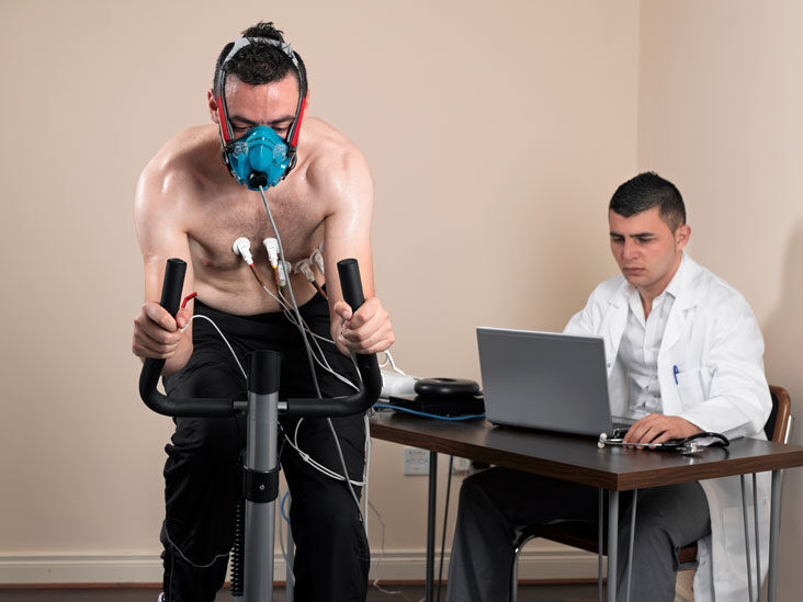
A doctor can ask for an echocardiogram as a part of a stress test.
Trusted supply involves physical exertion, like walking or cardiopulmonary exercise on a treadmill. Throughout the test to take a look at, monitor pulse, pressure, and also the heart’s electrical activity.
A sonographer can take a transthoracic echocardiogram before starting and ending the exercise:
Doctors use stress tests to diagnose:
- ischemic heart condition
- coronary heart condition
- heart failure
- problems causing the heart valves
Echocardiogram Risks & Side Effects

In general, the test carries some risk of serious complications or facet effects. However, individuals could feel some discomfort, and a few people will have an allergy to the contrast medium or anesthetic.
Summary:
Doctors use diagnostic procedures to diagnose issues that have an effect on the heart. Throughout the test, a doctor can identify how well a person’s heart pumps blood. Doctors can even use diagnostic procedures to see for signs of heart condition, like a weak muscle, blood clots within the heart, or poorly functioning heart valves.
A doctor may order an echocardiogram if someone shows symptoms of heart conditions, such as:
- shortness of breath
- leg swelling
- heart murmurs
- irregular heartbeat
- abnormal pressure
You can also read our other blog on- Teleradiology: Another revolution in Radiology?