


Let’s Know More About CT Scans, Usages & Works! : Have you ever wondered what that machine is that allows doctors and surgeons to get a detailed view of what’s going on within our bodies? It’s known as CT SCAN. Let’s take a look at what a CT scan is, how it works, and why it is required.
WHAT IS A CT SCAN?
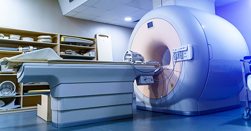
Computed tomography, usually known as a CT scan, is a technique used by doctors to evaluate inside body structures.
It can create images of a cross-section of your body by combining X-rays and computers. It takes photographs of your bones, muscles, organs, and blood arteries that display very thin “slices” of your body so that healthcare specialists may see your body in exquisite detail. A CT scan is essentially an X-ray examination in which a sequence of beams is rotated around a specific body component to obtain computer-generated cross-sectional images.
When compared to traditional X-rays, the advantage of tomographic images is that they contain comprehensive information on a specific area in cross-section, removing image superimposition, which gives them a significant advantage over plain films. For a suspected illness, it reveals an excellent clinicopathological correlation.
Low-dose CT scans are proving to be beneficial in cancer prevention and screening. The procedure was originally known as a CAT scan, which stood for computer axial tomography because the table moved after each axial image was taken.
NOW LET’S HAVE A LOOK AT HOW CT SCAN WORKS?

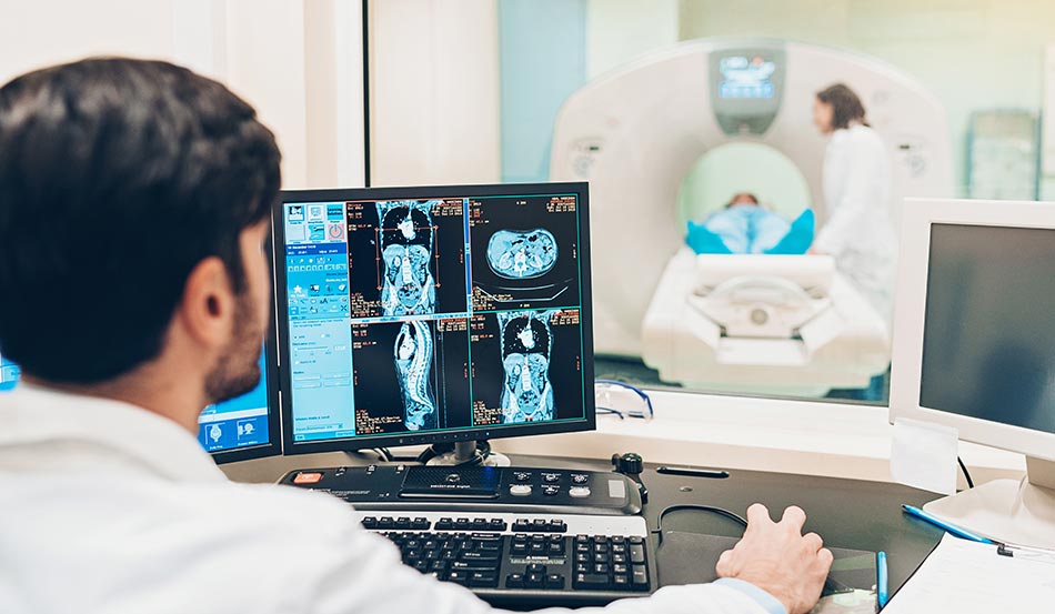
LOOK AT HOW CT SCAN WORKS: Through a circular frame known as the gantry, the CT scanner equipment revolves the X-ray tube around the patient’s body. Computerized data is acquired every time the machine rotates. Different cross-section photos are created while the subject is progressively moved up and down in the table. A 2D picture slice is created for each rotation.
The thickness of each consecutive picture slice is determined by the operator and the physician/radiologist but typically ranges from 1 to 10 millimeters. To accommodate the best cross-sectional image, the gantry can be shifted to the desired angle. A scan is converted into a computer image and can be simply replicated and stored once the necessary number of slices has been obtained. The image is formed using pixels that are radiosensitive and shown with Hounsfield scale units that are compared to known tissue density.
Water has a density of 0, air has a density of 1000, and bone has a density of 400 to 2000. Intravenous iodine can be administered into the bloodstream to identify infectious processes, blood arteries, and malignancies.

USAGES OF CT SCAN-
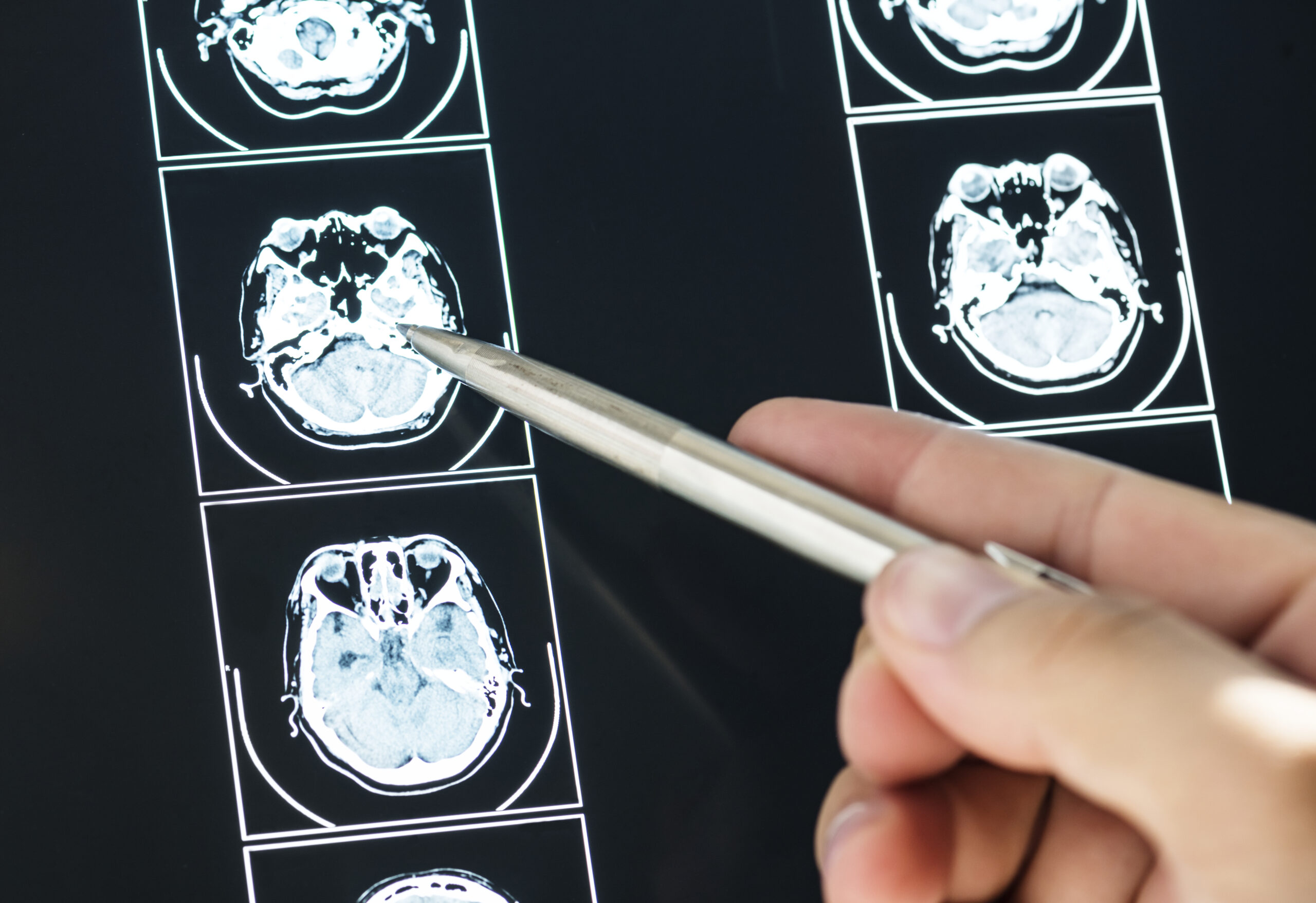
CT SCAN Uses: Depending on the organs to be evaluated, a CT scan is utilized for a variety of clinical indications. It can rule out serious sickness in an emergency situation. It is ordered to assist the physician in diagnosing, narrowing the differential diagnosis, and confirming the doctor’s assumptions.
Cancer screening, staging, and follow-up can all be done with it. Its use aids in performing proper biopsies and assisting during a surgical procedure.
Brain: tumors, traumatic or spontaneous hematomas, stroke, edema, skull fracture.
Neck: tumors, benign masses, thyroid nodules, lymphadenopathy
Chest: tumor, pneumonia, metastasis, benign masses.
Abdomen: primary tumors, metastases, abscess, ascites, cholecystitis.
Spine: fractures, degenerative changes, stability, osteomyelitis, disc pathology.
Bone: complex bone fractures, eroded joints, knee, tumors, osteomyelitis.
Gyn: cyst, fibromas, tumors
Screening: colon and lung cancer
Biopsy: guided to different organs for adequate tissue extraction
Angiography: brain, heart, lung, kidney, extremities
Intraoperative: it can be used for neuronavigation procedures during brain biopsy or tumor resection.

The estimated radiation collected from a single scan is similar to the person’s natural exposure to the environment for several months to a few years. If a patient is pregnant, the radiation dose is low and the fetus is normally unaffected.
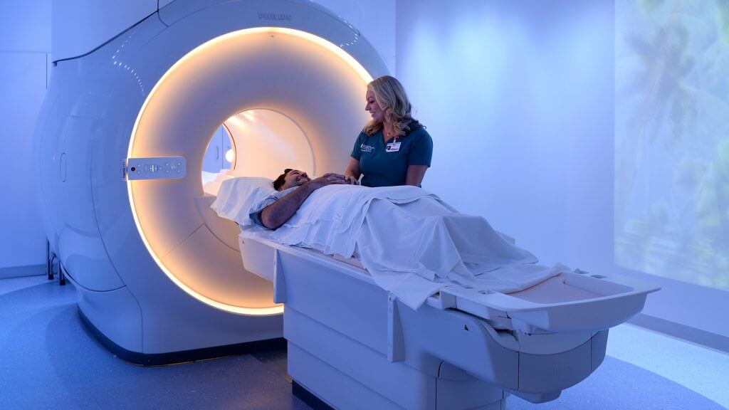
Recent Updates: Magnetic Resonance Imaging (MRI): Every essential detail you need to be aware off!!
Compared to magnetic resonance imaging, the CT scan has significant advantages. Because it has no effect on the device settings, it can be utilized safely in patients who have pacemakers, programmable pumps, or shunts. Patients who are claustrophobic normally cannot complete an MRI examination; nevertheless, they are more at ease during a CT scan because it is faster and quieter.